
Abstract
Chilaiditi’s sign, defined as the interposition of the colon’s hepatic flexure in front of the liver, is a rare condition. Although it is often asymptomatic, Chilaiditi’s sign can also present with symptoms and be associated with serious complications. A 65-year-old male patient diagnosed with chronic lymphocytic leukemia, considered as Rai stage 2, was admitted for observation and treatment arrangements following complaints of coughing. The air levels observed in the colonic loops between the liver and diaphragm on the posteroanterior chest radiographs were clinically and radiologically evaluated and presented as Chilaiditi’s sign.
Keywords: Chilaiditi’s sign, chronic lymphocytic leukemia, radiology
INTRODUCTION
Chilaiditi’s sign is characterized by the anomalous positioning of the right colon between the liver and the right hemidiaphragm (). Chilaiditi’s syndrome encompasses characteristic radiological findings along with symptoms such as nausea, vomiting, anorexia, constipation, and epigastric pain ().
While often asymptomatic and incidentally discovered, the Chilaiditi’s sign can be misinterpreted as conditions such as diaphragmatic hernia, subphrenic abscess, or pneumoperitoneum due to its rarity, despite the presence of characteristic air under the unchanged diaphragm resembling haustra (). Complications include perforation, colonic volvulus, internal hernia, subphrenic appendicitis, and acute intestinal obstruction (, ).
Management depends on the symptomatology. Asymptomatic cases generally require no treatment, whereas mild to moderate symptoms typically respond to conservative measures, such as cessation of oral intake, intravenous fluids, and bowel decompression. Persistent symptoms or complications such as ischemia or perforation necessitate surgical intervention (, ).
This case report highlights the potential for the Chilaiditi’s sign to present with non-specific symptoms and to mimic more serious conditions.
CASE REPORT
A 65-year-old male with untreated chronic lymphocytic leukemia (CLL), classified as stage 2 according to the Rai staging system, presented for advanced treatment (). His medical history includes a prior right inguinal hernia repair and current use of atorvastatin 10 mg/day. Family history is notable for Alzheimer’s disease in siblings and hypertension in a brother.
On examination, the patient appeared to be well-oriented and cooperative. Physical findings were unremarkable, except for splenomegaly that was palpable 6 cm below the costal margin. Respiratory examination revealed equally reduced aeration in both hemithoraces without adventitious sounds. There was no orthopnea.
Laboratory tests were performed (). The urinalysis was normal. The peripheral blood smear showed mature lymphocytes and basket cells without atypical findings.
Table 1. Laboratory test results of the patient.
Hemoglobin | 7.1 g/dL (12-16 g/dL) |
|---|---|
Hematocrit | 23.8% (40-54%) |
Leukocytes | 207.02x10³/µL (4-10x103/μL) |
Platelets | 140x103/µL (100-450x103/μL) |
Blood glucose | 141 mg/dL (70-105 mg/dL) |
Sodium | 140 mEq/L (136-145 mEq/L) |
Calcium | 7.5 mg/dL (8.4-10.2 mg/dL) |
Potassium | 4.2 mEq/L (3.5-5.1 mEq/L) |
Phosphorus | 3.7 mg/dL (2.3-4.7 mg/dL) |
Aspartate transaminase | 21 IU/L (5-34 IU/L) |
Alanine transaminase | 22 IU/L (<55) |
Uric acid | 5.8 mg/dL (2.6-6.0 mg/dL) |
Creatinine | 1.26 mg/dL (0.72-1.25 mg/dL) |
Alkaline phosphatase | 144 IU/L (40-150 IU/L) |
Total protein | 56.8 g/dL (64-83 g/dL) |
Albumin | 35.9 g/dL (35-50 g/dL) |
Blood urea nitrogen | 19.16 mg/dL (6-20 mg/dL) |
IU: International unit
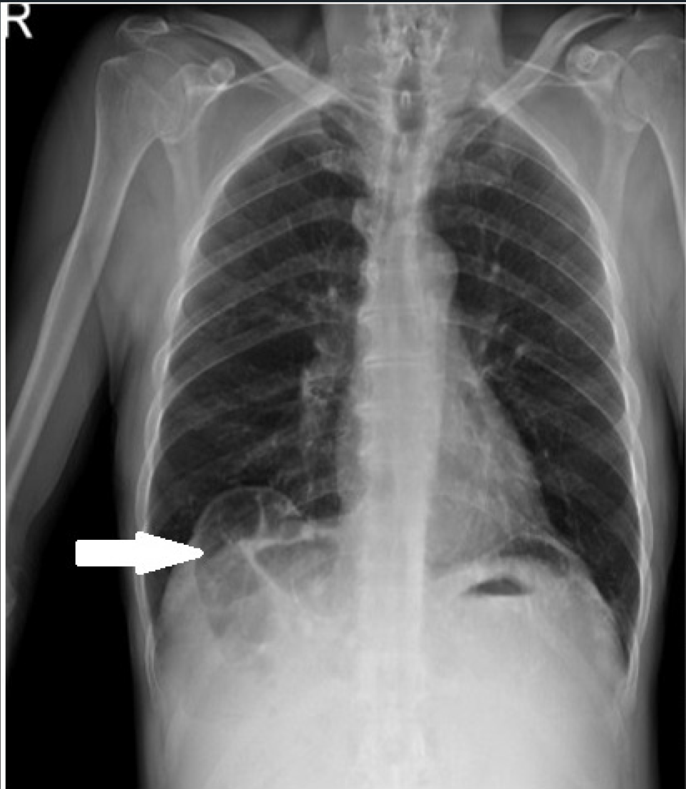
A pre-admission chest X-ray revealed elevation of the right hemidiaphragm with air between the right hemidiaphragm and liver ().
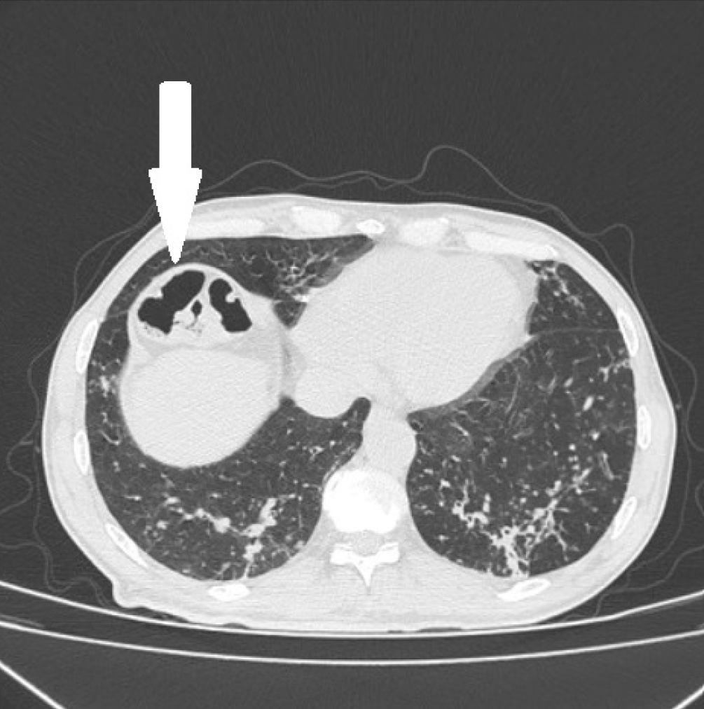
The presence of colonic haustral patterns was supported by thoracic computed tomography (CT) findings ().
CT: Computed tomography
Thoracic CT confirmed multiple parenchymal nodules with air bronchograms in the lower lobes of the lung and anterior positioning of the liver hepatic flexure.
During the hospitalization period, the patient remained asymptomatic for tumor lysis syndrome, with appropriate nutritional and treatment guidance provided in case symptoms developed.
DISCUSSION
Anomalous placement of the colon at the hepatic flexure, though rare, can range from asymptomatic to symptomatic with potential for serious complications, as demonstrated in this case (). Diagnosis in suspected cases, as illustrated here, can be straightforwardly confirmed by chest X-ray showing gas patterns crossing colonic haustral bands (). CT serves as a valuable tool for supportive and differential diagnosis. Management focuses on symptomatic relief with close monitoring for potential complications (). Given the absence of a documented association between CLL and Chilaiditi’s sign in literature, this case underscores the importance of recognizing and managing this condition independently.
Footnote
Ethics Committee Approval: N/A
Informed Consent: Informed consent was obtained from the patient.
Conflict of Interest: The authors declared no conflict of interest.
Author Contributions: Surgical and Medical Practices: A.Ş.A., İ.Y., Concept: A.Ş.A., İ.Y., Design: A.Ş.A., Data Collection and/or Processing: A.Ş.A., Analysis and/or Interpretation: A.Ş.A., İ.Y., Literature Search: A.Ş.A., Writing: A.Ş.A., İ.Y.
Financial Disclosure: The authors declared that this study received no financial support.
Footnotes
Ethics Committee Approval: N/A
Informed Consent: Informed consent was obtained from the patient.
Conflict of Interest: The authors declared no conflict of interest.
Author Contributions: Surgical and Medical Practices: A.Ş.A., İ.Y., Concept: A.Ş.A., İ.Y., Design: A.Ş.A., Data Collection and/or Processing: A.Ş.A., Analysis and/or Interpretation: A.Ş.A., İ.Y., Literature Search: A.Ş.A., Writing: A.Ş.A., İ.Y.
Financial Disclosure: The authors declared that this study received no financial support.
References
Sanyal K, Sabanathan K. Air below the right diaphragm: Chilaiditi sign. Emerg Med J. 2008;25(5):300-300.
Plorde JJ, Raker EJ. Transverse colon volvulus and associated Chilaiditi’s syndrome: case report and literature review. Am J Gastroenterol. 1996;91(12):2613-6.
Eisenstat TE, Raneri AJ, Mason GR. Volvulus of the transverse colon. Am J Surg. 1977;134(3):396-9.
Loke KL, Chan CS. Case report: transverse colon volvulus: unusual appearance on barium enema and review of the literature. Clin Radiol. 1995;50(5):342-4.
Sato M, Ishida H, Konno K et al. Chilaiditi syndrome: sonographic findings. Abdom Imaging. 2000;25(4):397-9.
Haddad CJ, Laclé J. Chilaiditi’s syndrome. A diagnostic challenge. Postgrad Med. 1991;89(4):249-50, 252.
Rai KR, Peterson BL, Bender N et al. The clinical staging of chronic lymphocytic leukemia. Blood. 1975;46(2):219-34.